


Endoscopy
Endoscopy is an advanced diagnostic method that allows for a thorough examination of the interior of the gastrointestinal tract using specialized equipment. Thanks to endoscopy in Białystok, doctors can precisely assess a patient's health, identifying any abnormalities. This procedure is invaluable in diagnosing numerous conditions, enabling quick and effective treatment.
Indications for endoscopic examinations
Endoscopy is recommended for symptoms such as chronic abdominal pain, difficulty swallowing, or gastrointestinal bleeding. This test is also recommended for individuals with suspected gastrointestinal cancer or inflammatory diseases. Regular endoscopy is crucial for individuals with a family history of gastrointestinal disease. Indications for endoscopy also include monitoring treatment progress and assessing the effectiveness of therapy. For patients with nonspecific gastrointestinal symptoms, endoscopy can help establish an accurate diagnosis.
What endoscopy tests do we perform?
The Gastroenterology Diagnostic Center in Białystok offers a wide range of endoscopic examinations, including gastroscopy, colonoscopy, and sigmoidoscopy. Each of these examinations allows for a detailed assessment of different sections of the gastrointestinal tract, which is crucial for making an accurate diagnosis. Endoscopic services also include biopsies and polyp removal, allowing for quick and effective intervention if abnormalities are detected.
Gastroscopy
What is this?
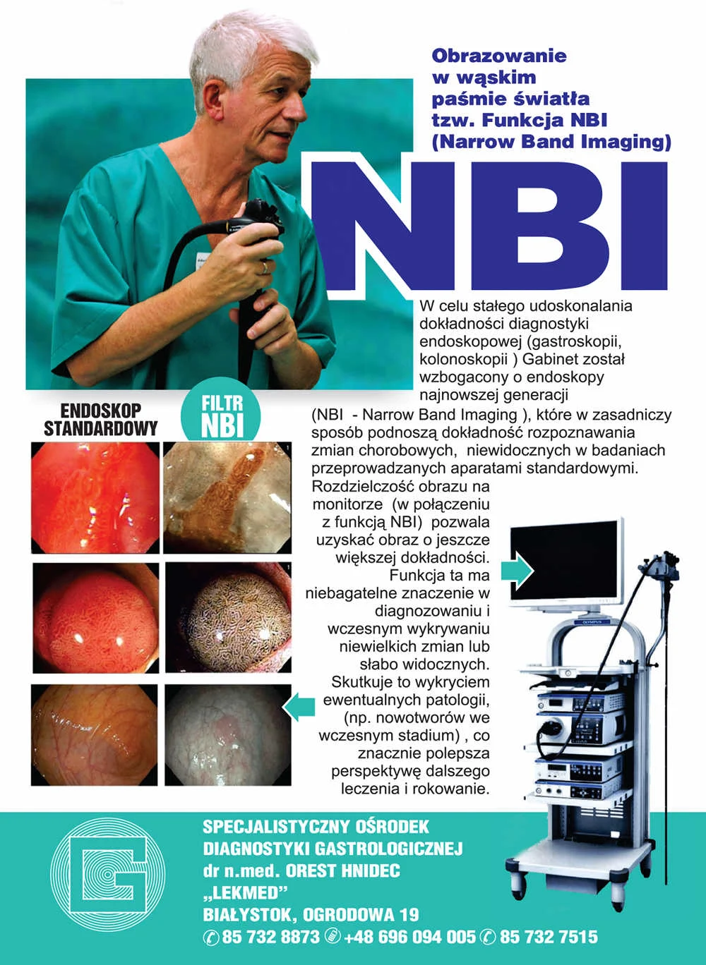
Gastroscopy is an endoscopic examination of the upper gastrointestinal tract, including the esophagus, stomach, and duodenum. The device used during the examination is called an endoscope. It consists of a flexible scope that allows for viewing the interior of the organ being examined. A microcamera with a wide field of view is placed at the end of the flexible scope, capturing a color image, which is then processed by a digital processor for easy viewing by the doctor. If necessary, forceps or a brush are inserted through the biopsy channel of the device to collect material for histopathological examination or for the presence of Helicobacter pylori bacteria, a so-called trauma test. Both the gastroscopy itself and the sampling for testing are completely painless procedures. The gag reflex may be a nuisance for the patient, but most patients cope well with it by breathing deeply and calmly.
For people with a very strong gag reflex, we recommend a very thin gastroscope that can be inserted into the stomach through the nose, avoiding irritation of the palate that causes gag reflexes. This is called STRESS-FREE GASTROSCOPY.
The purpose of gastroscopy
A gastroscopic examination allows the doctor to thoroughly assess the mucosa of the entire upper gastrointestinal tract. The elasticity and mobility of the esophageal, duodenal, and stomach walls, the visibility of blood vessels, folds, and peristaltic function are also assessed. Furthermore, the doctor pays attention to the amount and type of fluid content. Diagnostic gastroscopy is a safe procedure, so it can be performed both in the hospital and on an outpatient basis. The most common purpose of gastroscopy is to diagnose digestive system diseases:
- bacterial, viral, and fungal diseases
- exclusion of benign and malignant neoplastic lesions; In some cases, it is possible to detect early changes in:
- gastroesophageal reflux disease (GERD)
- inflammation of the mucosa of the esophagus, stomach, and duodenum
- searching for the site of bleeding
- searching for the cause of anemia
- gastric and duodenal ulcers
- changes caused by chemical factors: certain medications, acids and bases, bile
- during gastroscopy, it is possible to remove foreign bodies (swallowed or remaining after surgery, e.g., surgical threads), polyps (polypectomy), or flat lesions (mucosectomy), stop bleeding, band and obliterate esophageal varices
- assess the severity of disease changes and classify them accordingly, e.g., esophageal varices (present in portal hypertension - e.g., in liver cirrhosis), reflux esophagitis or sources of upper gastrointestinal bleeding. This assessment influences the treatment process.
When should you have a gastroscopy?
The indications are broad. The test should be performed in all individuals over 45 years of age with abdominal symptoms. In younger individuals, when there is a need to verify the diagnosis or alarm symptoms appear (weight loss, anemia, dysphagia, suspected gastrointestinal bleeding – bloody or coffee grounds-like vomiting, tarry or bloody stool). Additionally, if dyspeptic symptoms (pain, discomfort localized to the upper abdomen) lasting > 2 months occur, including in a person taking nonsteroidal anti-inflammatory drugs, unexplained chest pain, persistent symptoms suggestive of gastroesophageal reflux disease, nocturnal upper abdominal pain, and if celiac disease is suspected.
We perform screening or follow-up tests in patients at increased risk of developing cancer (people with long-term gastroesophageal reflux disease, Barrett's esophagus, pernicious anemia in the course of atrophic gastritis, after gastric resection)
If a gastric or duodenal ulcer is diagnosed, a traumatic test for Helicobacter pylori is recommended. A follow-up Helicobacter pylori eradication test should be performed at least 4 weeks after completing antibiotic therapy.
Contraindications to gastroscopy
Gastroscopy is a safe and short examination (usually a few minutes), minimally invasive, and painless. However, there are contraindications, including:
- Uncontrolled hypertension
- Hypotension and shock
- Recent myocardial infarction
- Acute respiratory failure
- Lack of patient cooperation
Pregnancy is not a contraindication to gastroscopy, but the need for it should be considered.
Preparing for a gastroscopy
If the examination is to be performed before 2 p.m., you should report to the endoscopy office on an empty stomach, if after 2 p.m. - you can eat a light meal, e.g. yogurt + a slice of bread, tea no later than 6 hours before the examination.
Who performs gastroscopy?
Gastroscopy is performed by an experienced doctor assisted by a nurse.
Other diagnostic methods
they cannot replace gastroscopy, they can be tests that complement the diagnosis and differentiate the causes of the symptoms.
Anesthesia (sedation)
To increase the comfort of the examination, 4 types of anesthesia can be used:
- Local anesthesia of the posterior pharynx with lidocaine spray (the most common method)
- Minimal sedation (intravenous, intramuscular, or oral administration of a sedative)
- Deeper sedation with analgesia (intravenous, intramuscular, and oral administration of an anesthetic and analgesic)
- Full anesthesia (performed by an anesthesiologist) with monitoring of basic physiological parameters (heart rate, respiration, blood pressure)
The course of the study
The patient lies on their left side, with their upper body slightly elevated. If they wear dentures, they should remove them before the examination. A plastic mouth guard is placed over the teeth/gums. The patient is then instructed to stick out their tongue, and the doctor inserts the gastroscope tip into their mouth and asks them to swallow. Guided by the endoscopic image, the endoscope is advanced through the esophagus into the stomach and duodenum.
Sigmoidoscopy
Sigmoidoscopy is an endoscopic examination of the large intestine. The principle of the examination is identical to that of a colonoscopy. The procedures differ in the scope of the bowel assessment – the left half of the large intestine is usually examined. Sigmoidoscopy is shorter, and patient preparation involves two enema sessions – one the evening before the procedure and the day of the examination. Sometimes, laxatives, similar to those used during colonoscopy, may be necessary. The appropriate preparation method is determined by a gastroenterologist.
Colonoscopy
What is this?
This is an endoscopic examination of the lower gastrointestinal tract (large intestine) using an endoscope, an instrument that allows for viewing the interior of the organ being examined. In colonoscopy, the endoscope is called a colonoscope. The flexible camera is inserted through the anus, slowly advanced through the rectum, and then along the entire large intestine to the end. If necessary, it is possible to examine the final section of the small intestine (part of the ileum). In state-of-the-art equipment, a microcamera with a wide field of view is placed at the end of the flexible camera, capturing a color image, which is then transmitted to a processor for digital processing. If necessary, forceps or a brush are inserted through the colonoscope's biopsy channel to collect material for histopathological examination. Collecting material for testing is painless.
The course of a colonoscopy examination
This is an endoscopic examination of the lower gastrointestinal tract (large intestine) using an endoscope, an instrument that allows for viewing the interior of the organ being examined. In colonoscopy, the endoscope is called a colonoscope. The flexible camera is inserted through the anus, slowly advanced through the rectum, and then along the entire large intestine to the end. If necessary, it is possible to examine the final section of the small intestine (part of the ileum). In state-of-the-art equipment, a microcamera with a wide field of view is placed at the end of the flexible camera, capturing a color image, which is then transmitted to a processor for digital processing. If necessary, forceps or a brush are inserted through the colonoscope's biopsy channel to collect material for histopathological examination. Collecting material for testing is painless.
The aim of the study is:
Endoscopic prevention, diagnosis, and treatment
Indications for colonoscopy include:
- Unexplained anemia
- Screening of the healthy population for polyps and cancer
- Blood in the stool or occult blood in the stool
- Suspected colon cancer
- Weight loss
- Inflammatory bowel disease - diagnosis and surveillance
- Loss of appetite without an identifiable cause
- Unexplained diarrhea
- Lower abdominal pain
- Irregular bowel movements
- Persistent urge to defecate or involuntary defecation
- Changed stool appearance (e.g., narrow, pencil-shaped stools)
Operative colonoscopy includes:
Removal of polyps, foreign bodies, widening of strictures, stopping bleeding.
A polyp is a descriptive term meaning any elevation of tissue above the mucosal surface. In the large intestine, we distinguish between cancerous and non-cancerous polyps. They can be single or multiple. The most common type of polyp in adults is cancerous polyp (adenomas). The likelihood of cancerous transformation depends on the type and size of the adenoma (tubular, tubulovillous, villous). Small adenomas rarely become malignant, while among polyps over 2 cm in diameter, almost half have features of invasive cancer. Determining the polyp type is a matter for the histopathologist. Colorectal adenomas can appear at any age, but a significant increase in incidence occurs in people over 30 years of age. The most common location is in the rectum and sigmoid colon. Polyps, as well as sometimes advanced colorectal tumors, usually cause no symptoms. However, if you experience symptoms (as described above) that may suggest an ongoing intestinal lesion, it's a good idea to perform a colonoscopy as a preventative measure and not ignore the telltale signs. All polyps up to 10 mm in diameter can be removed and should be removed during an outpatient diagnostic examination and then examined by a histopathologist to determine the type of polyp and the potential stage of the cancer. Endoscopic polyp removal is a completely painless procedure.
Preparing for a colonoscopy
There are several preparations available on the market to prepare patients for colonoscopy. In our experience, the most effective preparation is FORTRANS + ESPUMISAN EASY.
Description of preparation for the FORTRANS test
- From about a week before the examination, please do not eat stone fruit, especially those with small seeds (grapes, kiwi, strawberries, apples), because they remain in the intestine for a longer time and when trying to suction the remaining liquid content from the intestine, they may cause the device to become clogged during the examination, which in consequence sometimes makes it impossible to continue the examination.
- The intestine must be well cleansed, then the image obtained will be clear.
- 2 days before the test you should follow a liquid diet (e.g. blended soup).
- In 1 liter of boiled or non-carbonated mineral water, dissolve 1 sachet of Fortrans + 1 sachet of Espumisan Easy (totalling 4 liters of liquid). You can improve the taste by adding lemon or grapefruit juice to the solution.
- On the day before the examination, e.g. from 6 p.m., you should start drinking the prepared laxative solution, systematically sipping a glass every 15 minutes (2 liters), the remaining part should be drunk in the morning at the same pace, finishing drinking 4-6 hours before the examination date.
Taking medications while preparing for the test:
-
You should not take medications that inhibit blood clotting (Acard, Polocard, Polopiryna, etc.) for a week before the test.
-
If you have diabetes or are taking other anticoagulants (e.g., Sintrom), you should discuss your colonoscopy preparation with your treating physician (who may temporarily switch you to a different medication).
-
Regardless of the time of the test, you can take your regular medications (cardiac, antihypertensive, and others) with water in the morning.
Who performs a colonoscopy?
Colonoscopy should be performed by an experienced physician assisted by a nurse.
Anesthesia (sedation)
The following types of anesthesia can be used to enhance the comfort of the examination:
- Local anesthesia of the anal canal with lidocaine gel
- Minimal sedation (intravenous, intramuscular, or oral administration of a sedative)
- Deeper sedation with analgesia (intravenous, intramuscular, and oral administration of an anesthetic and analgesic)
- Full anesthesia (performed by an anesthesiologist) with monitoring of basic physiological parameters (heart rate, respiration, blood pressure)
MAKE AN APPOINTMENT OR CALL!
OUR CONSULTANTS WILL HELP YOU CHOOSE A CONVENIENT DATE FOR YOUR TEST OR CONSULTATION.